
Neurological complaints—like headaches, seizures, weakness, tremors, memory issues—can be daunting. Neurologists use a suite of diagnostic tools to precisely identify issues in the brain, spinal cord, peripheral nerves, or neuromuscular junction. With early and accurate testing, appropriate treatment plans are possible. This guide demystifies the key tools used in neurology, helping patients better understand the process and reduce anxiety.
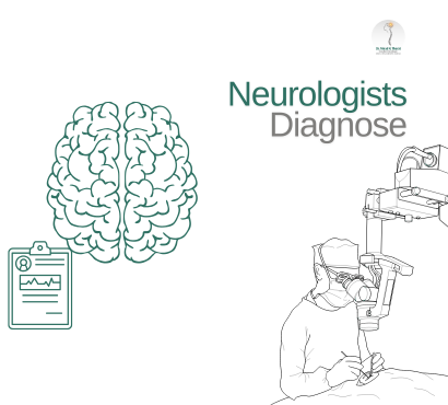
2 | Clinical Examination: The Starting Point
Every neurological diagnosis begins with a detailed history and physical exam, assessing:
- Symptom timeline, triggers, progression
- Motor signs: muscle strength, tone
- Reflexes pattern
- Coordination, gait, and balance
- Sensory function
- Cognitive and speech testing
- Cranial nerve examination
This guides decision-making toward specific tests for imaging, electrical studies, fluid analysis, or cognitive evaluation.
3 | Brain & Spinal Imaging
A. Magnetic Resonance Imaging (MRI)
- Uses magnets to produce detailed 3D images of brain and spinal cord.
- Detects stroke, tumors, demyelination (e.g., MS), infections.
- Contrast (gadolinium) may highlight inflammation.
- Takes 30–60 minutes; silent version, metal-free, open MRI options available.
B. Computed Tomography (CT)
- Uses X-rays to rapidly detect bleeding (stroke, trauma, hemorrhage).
- Quicker and more accessible; less detailed for soft tissue.
C. Functional MRI / MR Angiography
- Visualizes blood flow in the brain—useful for aneurysm and vascular evaluation.
4 | Electrical Activity Tests
A. Electroencephalogram (EEG)
- Measures brainwaves via scalp electrodes.
- Used for epilepsy, seizures, altered consciousness, encephalopathy.
- 20–60 minute outpatient test—regular or sleep-deprived EEGs provide deeper insight.
B. Evoked Potentials
- Measures nerve pathway function using stimuli:
- Visual (VEP), auditory (AEP), somatosensory (SEP).
- Detects latent demyelination (e.g., MS) and sensory pathway issues.
5 | Peripheral Nerve & Muscle Studies
A. Nerve Conduction Study (NCS)
- Measures electrical nerve speed—used for neuropathy, carpal tunnel, nerve injury.
B. Electromyography (EMG)
- Measures muscle electrical activity via needle electrodes.
- Diagnoses motor neuron disease, myopathy, nerve entrapment.
6 | Cerebrospinal Fluid Analysis (Lumbar Puncture)
- A small fluid sample tests for infection (meningitis), inflammation (neurosyphilis), demyelination, and bleeding.
- Usually 10 mL withdrawn from lower spine; short rest needed afterward.
7 | Cognitive & Neuropsychological Testing
- Detailed assessment in cases of dementia, memory loss, behavioral changes.
- Measures memory, attention, spatial skills—guides early detection and treatment areas.
8 | Emerging Tools
A. High-Resolution Ultrasound
- Visualizes nerve and muscle structure—useful in neuromuscular conditions.
B. MR Spectroscopy
- Checks chemical signatures of brain tissue—useful for tumors and metabolic disorders.
C. Wearable EEG/Remote Monitoring
- Ambulant seizure monitoring improving diagnostic accuracy.
9 | Choosing the Right Test
The neurologist’s choice depends on symptoms:
- Seizures → EEG ± MRI
- Sudden numbness/weakness → CT or MRI
- Neuropathy signs → NCS/EMG
- Memory decline → Cognitive testing + MRI
- Fever and encephalopathy → Lumbar puncture + imaging
10 | Patient Experience & Preparation
A. MRI/CT
- Comfortable clothing, removal of metal; may require sedation for those anxious or claustrophobic.
B. EEG/NCS/EMG
- No caffeine; painless but users may need to tolerate minor electrical sensations.
C. Lumbar Puncture
- Small needle, local numbness, patient lies flat for a few hours; rare side effects include headache.
11 | Interpreting Results & Next Steps
- Neurologist reviews findings, diagnoses condition
- Discusses treatments: medication, surgery, physiotherapy, rehabilitation
- Follow-up imaging or testing maps progress and response to treatment
Modern neurology uses a combination of clinical judgement and diagnostic precision tools—from advanced imaging to electrical and cognitive studies—to demystify complex brain and nerve disorders. Awareness of how tests are performed helps support patient confidence during the diagnostic journey. If you’re facing neurological symptoms, understanding these tools empowers you to make informed decisions and prepare effectively for the road ahead.
FAQ
Q1. What is better: MRI or CT scan?
MRI provides more detailed soft-tissue evaluation—ideal for stroke, tumors. CT is quicker, ideal for emergency bleeding and trauma.
Q2. Do EEGs hurt?
No—EEG is painless. Gel on the scalp may feel slightly cold; sleep-deprived tests may cause fatigue or drowsiness.
Q3. Is a lumbar puncture safe?
Yes. It’s performed under local anaesthetic, with low risk. Mild headache afterwards is common but usually short-lived with rest and hydration.