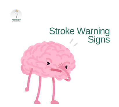
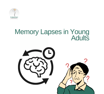
Dr Ninad Thorat

Category: Uncategorized

Uncategorized

Exercise-and-Brain-Health

Exercise and Brain Health: How Physical Activity Protects Your Nervous System

Physical activity is widely known for strengthening muscles and improving cardiovascular health, but its impact on the brain and nervous system is equally powerful. Research consistently shows that exercise and brain health are closely connected, with regular physical activity supporting memory, concentration, emotional balance, and long-term neurological protection. The nervous system controls everything from movement and breathing to thinking and emotions. Maintaining its health is essential for overall well-being. Regular exercise helps improve communication between brain cells, supports nerve regeneration, and reduces the risk of neurological diseases. Understanding how physical activity benefits brain function can encourage healthier lifestyle choices that protect cognitive abilities throughout life. Understanding the Nervous System and Its Role The nervous system consists of two main components: Central Nervous System (CNS) Includes: Brain Spinal cord Responsible for processing information and controlling body functions. Peripheral Nervous System (PNS) Includes nerves throughout the body that transmit signals between the brain and organs. Together, these systems regulate: Movement Memory Emotions Sensory perception Decision-making Reflex responses Maintaining nervous system health ensures efficient body functioning. How Exercise Improves Brain Function Regular physical activity strengthens brain performance in multiple ways. Increases Blood Flow to the Brain Exercise improves circulation, delivering: Oxygen Glucose Nutrients These are essential for brain cell survival and function. Stimulates Growth of New Brain Cells Exercise promotes neurogenesis, particularly in the hippocampus—the brain region responsible for memory and learning. Enhances Neurotransmitter Activity Physical activity improves production of: Dopamine Serotonin Endorphins These chemicals regulate mood, motivation, and mental clarity. Exercise Helps Improve Memory and Learning Ability Memory decline can occur with aging, stress, or sedentary lifestyles. Exercise supports memory retention by strengthening neural pathways. Benefits include: Better concentration Faster learning ability Improved decision-making Stronger recall capacity Regular physical activity is especially beneficial for students, professionals, and older adults. Role of Exercise in Preventing Neurological Disorders Physical activity significantly lowers the risk of several neurological conditions. These include: Stroke Exercise improves blood circulation and reduces blood pressure. Alzheimer’s Disease Regular activity supports brain plasticity and slows cognitive decline. Parkinson’s Disease Exercise improves coordination and movement control. Peripheral Neuropathy Movement helps maintain nerve strength and function. Consistent physical activity supports long-term neurological protection. Exercise Reduces Stress and Supports Mental Health Stress negatively affects nervous system performance. Chronic stress increases cortisol levels, which can damage brain cells over time. Exercise helps by: Reducing anxiety levels Improving mood stability Enhancing emotional resilience Promoting relaxation Supporting better sleep quality Mental health and neurological health are deeply interconnected. Physical Activity Improves Coordination and Balance Balance and coordination depend on healthy nerve communication between the brain and muscles. Exercise strengthens this connection through: Muscle activation Joint stability Reflex response improvement Activities like yoga and balance training are especially beneficial. Best Types of Exercise for Brain Health Different forms of exercise contribute to nervous system protection. Aerobic Exercise Examples include: Walking Jogging Cycling Swimming These improve blood flow to the brain. Strength Training Supports muscle-to-brain communication and improves coordination. Yoga Enhances relaxation and improves nerve signaling. Stretching Exercises Maintain flexibility and prevent nerve compression. Balance Training Supports reflexes and prevents falls, especially in older adults. Combining multiple exercise types produces the best neurological benefits. Exercise Supports Healthy Aging of the Brain Aging naturally affects cognitive function. However, regular physical activity slows this process significantly. Benefits for older adults include: Improved memory Reduced dementia risk Better reaction time Stronger emotional health Improved independence Exercise acts as a protective shield for aging brain cells. How Exercise Improves Sleep Quality Sleep is essential for nervous system repair and memory consolidation. Regular physical activity: Regulates sleep cycles Reduces insomnia Improvest sleep depth Enhances recovery of brain cells Better sleep directly supports neurological performance. Role of Exercise in Enhancing Focus and Productivity Physical activity improves mental clarity and concentration. Professionals and students often notice: Faster thinking ability Improved task completion speed Increased creativity Better decision-making skills Exercise strengthens the brain’s executive functioning ability. Exercise and Emotional Stability Mood disorders often affect nervous system balance. Physical activity supports emotional regulation through hormone balance. Benefits include: Reduced depression symptoms Improved confidence Greater emotional control Better stress tolerance Exercise acts as a natural mood stabilizer. How Much Exercise Is Needed for Brain Health? Health experts recommend: Adults At least 150 minutes of moderate exercise weekly or 75 minutes of vigorous exercise weekly Children and Adolescents Minimum 60 minutes daily physical activity Even small lifestyle changes like walking daily can improve neurological health. Warning Signs of Poor Nervous System Health Consult a neurologist doctor if symptoms persist such as: Frequent headaches Memory loss Tingling sensations Poor coordination Persistent fatigue Sleep disturbances Mood instability Early intervention prevents long-term neurological complications. Lifestyle Habits That Strengthen Brain Function Along With Exercise Exercise works best when combined with healthy daily habits. Important supportive habits include: Balanced nutrition Adequate hydration Stress management Quality sleep Mental stimulation activities Avoiding smoking Limiting alcohol consumption These habits enhance exercise-related neurological benefits. Long-Term Benefits of Exercise on the Nervous System Regular physical activity provides lasting protection against neurological decline. Key long-term advantages include: Improved cognitive performance Stronger nerve signaling Reduced dementia risk Better emotional health Enhanced learning ability Faster reflex responses Exercise supports both mental and physical independence throughout life. FAQ 1. Can exercise improve memory power? Yes. Regular physical activity increases blood flow to memory-related brain areas and supports growth of new neurons. 2. Which exercise is best for brain health? Aerobic exercises such as walking, cycling, and swimming are highly beneficial, especially when combined with strength training and yoga. 3. How quickly does exercise affect brain function? Some benefits like mood improvement appear immediately, while memory and cognitive improvements develop over weeks of regular activity.

Exercise and Brain Health: How Physical Activity Protects Your Nervous System Read More »

Amyotrophic-Lateral-Sclerosis

ALS (Amyotrophic Lateral Sclerosis): What You Need to Know

ALS (Amyotrophic Lateral Sclerosis) is a progressive neurological disorder that affects the nerve cells responsible for controlling voluntary muscle movement. It belongs to a group of conditions known as motor neuron diseases, where communication between the brain and muscles gradually weakens over time. ALS leads to muscle weakness, loss of movement control, and eventually difficulty with speaking, swallowing, and breathing. While the disease currently has no cure, early diagnosis and supportive treatment can significantly improve quality of life and slow progression in some cases. Understanding ALS symptoms, risk factors, and available treatment options helps patients and families manage the condition more effectively. What Is ALS (Amyotrophic Lateral Sclerosis)? ALS is a condition in which motor neurons, the nerve cells responsible for muscle movement, gradually degenerate and stop functioning. Motor neurons include: Upper motor neurons (brain to spinal cord) Lower motor neurons (spinal cord to muscles) As these neurons deteriorate: Muscles weaken Movement becomes difficult Coordination declines Muscle wasting develops Eventually, the body loses the ability to perform voluntary movements. Why ALS Is Also Called Motor Neuron Disease ALS is one of the most common forms of motor neuron disease (MND). The term describes disorders affecting: Muscle movement control Speech Swallowing Breathing ALS specifically affects both upper and lower motor neurons, which leads to progressive muscle weakness throughout the body. Early Symptoms of ALS Early symptoms of ALS may be subtle and often mistaken for general fatigue or muscle strain. Common early warning signs include: Muscle Weakness Usually begins in: Hands Legs Arms Feet Patients may notice difficulty holding objects or climbing stairs. Muscle Twitching (Fasciculations) Involuntary muscle movements are common early indicators. Slurred Speech Speech may become unclear due to weakened facial muscles. Difficulty Swallowing Some individuals experience choking while eating. Frequent Tripping or Falling Balance and coordination gradually decline. Recognizing these symptoms early supports timely neurological evaluation. Advanced Symptoms of ALS As ALS progresses, symptoms become more severe and widespread. These include: Severe muscle weakness Muscle wasting Difficulty walking Loss of hand function Breathing difficulty Speech impairment Swallowing problems Eventually, patients require assistance with daily activities. Causes of ALS The exact cause of ALS is not fully understood. However, several factors may contribute to disease development. Genetic Factors Approximately 5–10% of ALS cases are inherited. These are known as familial ALS. Environmental Factors Possible triggers include: Exposure to toxins Heavy metals Air pollution Agricultural chemicals Research is ongoing to confirm these links. Age ALS is more commonly diagnosed between ages 40 and 70. Gender Men are slightly more likely to develop ALS than women. Risk Factors Associated With ALS Although anyone can develop ALS, certain risk factors increase likelihood. These include: Family history of ALS Smoking Military service exposure risks Occupational toxin exposure Increasing age Understanding risk factors supports early medical monitoring. How ALS Affects the Nervous System ALS damages motor neurons that carry signals from the brain to muscles. As communication weakens: Muscles stop responding properly Movement becomes restricted Muscle tissue shrinks over time Importantly, ALS usually does not affect intelligence, memory, or senses in most patients. However, some individuals may experience mild cognitive changes. How ALS Is Diagnosed Diagnosing ALS can be challenging because symptoms resemble other neurological conditions. Doctors typically perform multiple evaluations. Neurological Examination Checks muscle strength, reflexes, and coordination. Electromyography (EMG) Measures electrical activity in muscles. Helps detect nerve damage. Nerve Conduction Studies Assess signal transmission between nerves and muscles. MRI Scan Used to rule out other neurological disorders. Blood and Urine Tests Help exclude alternative causes of symptoms. Diagnosis is based on combined clinical findings rather than a single test. Stages of ALS Progression ALS progression varies among individuals but generally follows predictable stages. Early Stage Symptoms include: Muscle weakness Twitching Mild coordination problems Daily activities remain mostly independent. Middle Stage Symptoms expand to multiple muscle groups. Patients may experience: Walking difficulty Speech changes Swallowing problems Supportive devices may become necessary. Late Stage Severe muscle weakness develops. Patients may experience: Limited movement Breathing difficulties Need for assisted ventilation Supportive care becomes essential. Treatment Options for ALS Although ALS currently has no cure, treatment focuses on slowing progression and improving quality of life. Medications Certain medications may: Slow disease progression Reduce nerve damage Manage symptoms Physical Therapy Helps maintain muscle strength and flexibility. Prevents joint stiffness. Speech Therapy Supports communication ability as speech weakens. Occupational Therapy Assists patients in adapting daily routines. Improves independence. Respiratory Support Breathing assistance may be required in later stages. Multidisciplinary care improves overall patient outcomes. Importance of Nutritional Support in ALS Maintaining proper nutrition becomes difficult as swallowing muscles weaken. Balanced nutrition helps: Maintain body weight Improve energy levels Support immune function Dietary adjustments are often recommended by specialists. Role of Assistive Devices in ALS Care Supportive devices improve mobility and independence. Common examples include: Walking aids Wheelchairs Communication devices Feeding assistance tools Breathing support equipment These technologies significantly enhance quality of life. Emotional and Psychological Impact of ALS Living with ALS affects both physical and emotional health. Patients may experience: Anxiety Depression Frustration Social isolation Mental health support plays an important role in overall care management. Family support also contributes to improved coping ability. Can ALS Be Prevented? Currently, there is no proven way to prevent ALS. However, maintaining general neurological health may reduce overall risk. Healthy lifestyle habits include: Regular physical activity Avoiding smoking Reducing toxin exposure Eating balanced nutrition Managing chronic diseases Early neurological consultation improves management outcomes. When Should You Consult a Doctor? Medical evaluation is recommended if symptoms appear such as: Persistent muscle weakness Muscle twitching Difficulty speaking Trouble swallowing Loss of coordination Frequent falls Early neurological assessment helps rule out serious conditions. FAQ 1. Is ALS a hereditary disease? Only about 5–10% of ALS cases are inherited. Most cases occur without a family history. 2. Does ALS affect thinking ability? Most patients retain normal intelligence, although mild cognitive changes can occur in some individuals. 3. Can ALS be cured? Currently, there is no cure for ALS, but treatment helps slow progression and

ALS (Amyotrophic Lateral Sclerosis): What You Need to Know Read More »

Dystonia

Dystonia: Uncontrollable Muscle Contractions and Treatment Breakthroughs

Movement is something most people rarely think about. From writing and walking to speaking and blinking, the body performs thousands of coordinated movements every day without conscious effort. However, for individuals living with dystonia, these everyday actions can become difficult and unpredictable. Dystonia is a neurological movement disorder characterized by involuntary muscle contractions that cause repetitive movements, abnormal postures, or twisting motions. These contractions can affect a single muscle, a group of muscles, or sometimes the entire body. Although dystonia may not be as widely recognized as other neurological disorders, it affects people of all ages and can significantly impact daily life. The condition may interfere with activities such as writing, walking, speaking, or maintaining normal posture. In recent years, research has improved our understanding of dystonia and led to several advances in treatment options. While dystonia cannot always be cured, modern therapies and medical innovations have made it increasingly manageable. This article explores what dystonia is, why it occurs, how it affects the body, how it is diagnosed, and the latest treatment breakthroughs that are improving the lives of individuals living with this condition. Understanding Dystonia Dystonia is a disorder that affects the way the brain controls muscle movements. Normally, the brain sends carefully coordinated signals to muscles, allowing them to contract and relax in a balanced way. In dystonia, these signals become disrupted. As a result, muscles contract involuntarily and may remain in that contracted state longer than normal. These abnormal contractions can cause: Twisting movements Abnormal postures Tremors Muscle stiffness Difficulty performing precise tasks The severity of dystonia varies from mild to severe. Some individuals may experience occasional muscle spasms, while others may struggle with persistent muscle contractions that affect mobility and daily activities. Types of Dystonia Dystonia is classified in several ways depending on the muscles involved and the underlying cause. Focal Dystonia Focal dystonia affects a single part of the body. Common examples include: Neck (cervical dystonia) Eyelids (blepharospasm) Hands or fingers (writer’s cramp) This is one of the most common forms of dystonia. Segmental Dystonia Segmental dystonia affects two or more neighboring body regions. For example, the neck and shoulders may be involved together. Generalized Dystonia In generalized dystonia, multiple parts of the body are affected, sometimes including the trunk and legs. This form is more commonly seen in younger individuals. Task-Specific Dystonia This form occurs only during certain activities. Examples include: Writer’s cramp during writing Musician’s dystonia during instrument performance The muscles behave normally during other tasks. Secondary Dystonia Secondary dystonia develops as a result of another underlying condition, such as brain injury, infection, medication side effects, or neurological disorders. What Causes Dystonia? The exact cause of dystonia is not always clear. However, scientists believe that it involves dysfunction in specific brain regions responsible for movement control. The basal ganglia, a group of structures deep inside the brain, play a crucial role in coordinating muscle activity. When signals within this system become disrupted, abnormal muscle contractions can occur. Several factors may contribute to the development of dystonia. Genetic Factors Some forms of dystonia are inherited. Certain genetic mutations may alter the way nerve cells communicate, affecting muscle control. Genetic dystonia often appears during childhood or adolescence. Brain Injury or Trauma Damage to the brain caused by injury, stroke, or oxygen deprivation may disrupt movement control pathways. Medication Effects Certain medications, particularly those affecting dopamine signaling in the brain, may trigger dystonic reactions. Neurological Disorders Dystonia may occur alongside other neurological conditions that affect movement control. Environmental Factors Although less common, environmental exposures or infections affecting the nervous system may also contribute to dystonia. Symptoms of Dystonia Symptoms of dystonia vary widely depending on which muscles are involved. Common signs include: Involuntary Muscle Contractions Muscles contract without voluntary control, often repeatedly. Abnormal Postures Affected body parts may twist or bend into unusual positions. Tremors Some individuals experience shaking movements similar to tremors. Muscle Pain or Fatigue Continuous muscle contractions may lead to discomfort or fatigue. Worsening With Activity Symptoms often become more noticeable during voluntary movement or stress. Improvement During Rest In some cases, symptoms may reduce when the body is relaxed or sleeping. How Dystonia Is Diagnosed Diagnosing dystonia requires a detailed evaluation of symptoms and medical history. Because dystonia symptoms may resemble other movement disorders, accurate diagnosis is important for proper treatment. Doctors typically assess: Symptom patterns Duration and progression Family history Neurological examination findings Additional tests may include imaging or laboratory tests to rule out other neurological conditions. How Dystonia Affects Daily Life Dystonia can influence many aspects of everyday living depending on its severity and location. Challenges may include: Difficulty writing or typing Problems with walking or balance Speech difficulties Eye closure affecting vision Social discomfort due to visible muscle movements Living with dystonia can also lead to emotional stress, frustration, and reduced confidence. Support and proper treatment play an important role in improving quality of life. Treatment Options for Dystonia Although dystonia cannot always be completely cured, several treatments can help control symptoms and improve daily functioning. Treatment plans are typically individualized based on the type and severity of dystonia. Medications Certain medications help regulate abnormal muscle activity by influencing neurotransmitters in the brain. These medications may reduce muscle spasms and improve movement control. Botulinum Toxin Injections Botulinum toxin injections are widely used to treat focal dystonia. These injections temporarily weaken overactive muscles, reducing involuntary contractions and improving posture or movement. The effects typically last several months and may require repeat treatments. Physical Therapy Physical therapy plays an important role in managing dystonia. Therapeutic exercises may help: Improve flexibility Reduce muscle stiffness Enhance posture Strengthen unaffected muscles Stretching and relaxation techniques may also help relieve muscle tension. Occupational Therapy Occupational therapy focuses on helping individuals perform daily tasks more comfortably. Therapists may recommend adaptive techniques or tools that make activities such as writing, eating, or working easier. Deep Brain Stimulation (DBS) Deep brain stimulation is one of the most significant breakthroughs in dystonia treatment. This procedure involves implanting

Dystonia: Uncontrollable Muscle Contractions and Treatment Breakthroughs Read More »

neurologist-in-nashik

Essential Tremor: When Shaking Interferes With Daily Life

Occasional shaking or trembling of the hands is something most people experience at some point in their lives. It can happen due to stress, fatigue, anxiety, or even excessive caffeine intake. However, when shaking becomes persistent and begins to interfere with daily activities such as writing, eating, or holding objects, it may indicate a neurological condition known as essential tremor. Essential tremor is one of the most common movement disorders affecting millions of people worldwide. Although it is not life-threatening, the condition can significantly affect quality of life, especially when symptoms worsen over time. Many individuals initially ignore the symptoms or assume they are simply a normal part of aging, which often delays diagnosis and management. Understanding essential tremor is important because early recognition and appropriate treatment can help reduce symptoms and improve daily functioning. This article explores what essential tremor is, its causes, symptoms, diagnosis, treatment options, and lifestyle strategies that help individuals manage the condition effectively. What Is Essential Tremor? Essential tremor is a neurological disorder that causes involuntary and rhythmic shaking, most commonly affecting the hands. However, tremors may also occur in other parts of the body, including the head, voice, arms, and occasionally the legs. Unlike some other neurological disorders, essential tremor is primarily characterized by action tremors, meaning the shaking becomes noticeable when a person tries to perform a task. For example, tremors may become more visible when holding a cup, writing, or using utensils. The condition may develop gradually and typically progresses slowly over time. While it can occur at any age, it is more commonly observed in adults over the age of 40. How Essential Tremor Affects Daily Life Although essential tremor is not usually dangerous, it can interfere with routine activities. People with moderate to severe tremors may struggle with: Drinking from a glass without spilling Writing clearly Using tools or utensils Applying makeup or shaving Holding small objects In some cases, tremors affecting the voice may cause shaky speech, which can affect communication and confidence in social situations. These challenges can lead to frustration, embarrassment, and reduced independence if the condition is not managed properly. Causes of Essential Tremor The exact cause of essential tremor is not fully understood, but researchers believe it is related to abnormal communication between certain areas of the brain that control movement. Several factors may contribute to the condition. Genetic Factors Essential tremor often runs in families. In many cases, a person with essential tremor has a parent or close relative who also experiences similar symptoms. This hereditary form is sometimes referred to as familial tremor. Brain Signal Abnormalities Some studies suggest that changes in brain circuits involving the cerebellum—an area responsible for coordination and balance—may lead to tremor development. Age-Related Changes Although essential tremor can appear at any age, it becomes more common with advancing age. Common Symptoms of Essential Tremor The symptoms of essential tremor can vary from mild to severe and may gradually worsen over time. Hand Tremors The most common symptom is shaking of the hands, especially during activities such as writing, eating, or holding objects. Head Tremors Some individuals experience involuntary head movements that may appear as nodding or shaking from side to side. Voice Tremors Tremors affecting the vocal cords can lead to shaky or quivering speech. Arm or Leg Tremors Although less common, tremors may occasionally affect other parts of the body. Worsening with Activity Unlike tremors that occur at rest, essential tremor usually becomes more noticeable when performing tasks. Triggers That May Worsen Tremors Certain factors may temporarily increase tremor intensity. These include: Emotional stress or anxiety Fatigue Caffeine consumption Lack of sleep Certain medications Understanding these triggers can help individuals manage symptoms more effectively. Essential Tremor vs Parkinson’s Disease Essential tremor is sometimes confused with Parkinson’s disease because both conditions involve shaking. However, they are different disorders with distinct characteristics. Essential Tremor Occurs mainly during movement Often affects both sides of the body Usually involves the hands, head, or voice Parkinson’s Disease Tremors often occur at rest May start on one side of the body Associated with additional symptoms such as stiffness, slow movement, and balance problems A Neurologist evaluation is important to distinguish between the two conditions. How Essential Tremor Is Diagnosed There is no single test specifically designed to diagnose essential tremor. Instead, doctors rely on clinical evaluation and medical history. The diagnostic process may include: Physical and Neurological Examination The doctor evaluates tremor patterns, muscle coordination, and reflexes. Medical History Review Information about symptom onset, family history, and potential triggers helps guide diagnosis. Movement Observation Patients may be asked to perform certain tasks such as writing or holding objects to assess tremor severity. Additional Tests In some cases, imaging tests or blood tests may be performed to rule out other conditions that could cause tremors. Treatment Options for Essential Tremor Although essential tremor cannot always be completely cured, several treatment options can help manage symptoms and improve daily functioning. Medications Doctors may prescribe medications that help reduce tremor intensity. These medications work by influencing nerve signals involved in muscle movement. Not all patients respond to the same medications, so treatment plans may vary. Physical and Occupational Therapy Therapy can help individuals develop techniques to improve coordination and reduce tremor-related difficulties. Examples include: Strengthening exercises Hand control training Adaptive techniques for daily tasks Assistive Devices Certain devices may help people perform routine tasks more easily. These may include: Weighted utensils Stabilizing wrist supports Special writing tools Such tools can significantly improve independence and comfort. Surgical Treatment In severe cases where medications are not effective, surgical options may be considered. Advanced procedures target specific areas of the brain responsible for tremors and can help reduce symptoms. Surgical treatment is usually reserved for individuals whose tremors significantly affect daily life. Lifestyle Changes That Help Manage Tremors Lifestyle modifications can play an important role in controlling essential tremor symptoms. Reduce Caffeine Intake Caffeine stimulates the nervous system and may increase tremor severity. Manage Stress Stress and anxiety

Essential Tremor: When Shaking Interferes With Daily Life Read More »

trigeminal-neuralgia

Trigeminal Neuralgia: The “Suicide Disease” and Modern Management Options

Imagine experiencing sudden, electric shock-like pain on your face while doing something as simple as brushing your teeth, talking, or even feeling a light breeze. This is the harsh reality for individuals suffering from trigeminal neuralgia, a condition often referred to as the “suicide disease” due to the intensity and unpredictability of its pain. Despite being relatively rare, trigeminal neuralgia can severely impact a person’s quality of life—physically, emotionally, and socially. The good news is that modern medicine offers multiple treatment options that can significantly reduce pain and restore normalcy. In this comprehensive guide, we’ll explore everything you need to know about trigeminal neuralgia—from causes and symptoms to the latest management options available today. What is Trigeminal Neuralgia? Trigeminal neuralgia is a chronic pain condition affecting the trigeminal nerve, which is responsible for carrying sensory information from your face to your brain. Key Facts: It typically affects one side of the face Pain episodes are sudden and intense It can last for seconds to minutes but may recur frequently The trigeminal nerve has three branches: Ophthalmic (forehead and eyes) Maxillary (cheeks and upper jaw) Mandibular (lower jaw) Pain can occur in one or more of these regions. Why is it Called the “Suicide Disease”? The nickname comes from the extreme severity of pain associated with the condition. Many patients describe it as: Electric shock-like Burning or stabbing sensation Sudden and unbearable In severe, untreated cases, the pain can lead to: Depression Anxiety Social withdrawal Difficulty eating or speaking This highlights the importance of early diagnosis and proper treatment. Causes of Trigeminal Neuralgia Trigeminal neuralgia occurs when the trigeminal nerve is irritated or compressed. Common Causes: 1. Blood Vessel Compression A blood vessel pressing against the nerve is the most common cause. This leads to damage of the nerve’s protective covering (myelin sheath). 2. Age-Related Changes It is more common in individuals above 50 years of age due to natural wear and tear. 3. Multiple Sclerosis In some cases, nerve damage is linked to neurological conditions. 4. Tumors or Structural Issues Rarely, a tumor or abnormality may compress the nerve. 5. Unknown Causes (Idiopathic) In some patients, no clear cause is identified. Symptoms of Trigeminal Neuralgia The hallmark symptom is severe facial pain, but it presents in specific patterns. Typical Symptoms: Sudden, sharp, electric shock-like pain Episodes lasting a few seconds to minutes Pain triggered by simple activities like: Brushing teeth Talking Eating Touching the face Pain occurring in cycles (active phases followed by remission) Trigger Zones: Certain areas on the face can trigger pain when touched. Types of Trigeminal Neuralgia 1. Type 1 (Classic TN) Sudden, intense, intermittent pain Most common form 2. Type 2 (Atypical TN) Continuous dull aching pain Less intense but more persistent Diagnosis: How is Trigeminal Neuralgia Identified? Diagnosis is primarily based on clinical history and symptom patterns. Evaluation Includes: Detailed patient history Neurological examination MRI scan to rule out: Tumors Multiple sclerosis Vascular compression Early Neurological diagnosis is crucial to prevent worsening of symptoms. Modern Management Options Treatment depends on the severity, cause, and patient’s overall health. Modern management combines medications, minimally invasive procedures, and surgical options. 1. Medical Management (First-Line Treatment) Medications are usually the first step. Commonly Used Drugs: Anticonvulsants (to stabilize nerve activity) Muscle relaxants Pain modulators Benefits: Effective in early stages Non-invasive Limitations: Side effects like dizziness, fatigue Reduced effectiveness over time 2. Minimally Invasive Procedures When medications fail, minimally invasive treatments are considered. a) Radiofrequency Ablation Uses heat to damage pain-causing nerve fibers Provides quick pain relief b) Balloon Compression Compresses the nerve to block pain signals c) Glycerol Injection Chemical injection to reduce nerve pain Advantages: Quick recovery Day-care procedures Suitable for elderly patients Limitations: Temporary relief in some cases May require repeat procedures 3. Surgical Treatment For severe or resistant cases, surgery offers long-term relief. Microvascular Decompression (MVD) This is considered the most effective surgical option. Procedure: The surgeon relieves pressure on the nerve by repositioning the blood vessel Benefits: Long-lasting relief Preserves nerve function Risks: Requires general anesthesia Small risk of complications Lifestyle and Supportive Measures While medical treatment is essential, lifestyle adjustments can help manage symptoms. Practical Tips: Avoid known triggers (cold wind, hard foods) Maintain oral hygiene gently Eat soft foods during flare-ups Manage stress through relaxation techniques Emotional and Psychological Impact Living with trigeminal neuralgia is not just physically painful—it also affects mental health. Common Emotional Challenges: Fear of pain recurrence Anxiety during daily activities Social isolation Coping Strategies: Psychological counseling Support groups Family support Mindfulness and relaxation techniques Importance of Early Treatment Delaying treatment can lead to: Increased frequency of pain episodes Reduced response to medications Emotional distress Early intervention improves outcomes and quality of life. Advances in Treatment Modern advancements have significantly improved management: Improved imaging for accurate diagnosis Safer surgical techniques Better pain control medications Minimally invasive procedures with faster recovery When Should You See a Doctor? Seek medical help if you experience: Recurrent facial pain Sudden electric shock-like sensations Pain triggered by normal activities Persistent discomfort affecting daily life Early neurologist consultation can prevent complications. Trigeminal neuralgia may be one of the most painful medical conditions, but it is manageable with the right approach. From medications to advanced surgical options, modern treatments offer hope and relief to patients suffering from this condition. If you or someone you know is experiencing symptoms, don’t ignore them. Early diagnosis and proper care can make a life-changing difference. FAQs 1. Is trigeminal neuralgia a lifelong condition? It can be chronic, but many patients experience periods of remission. With proper treatment, symptoms can be effectively controlled and quality of life improved. 2. Can trigeminal neuralgia go away on its own? In some cases, symptoms may temporarily disappear, but the condition usually requires medical treatment to prevent recurrence and worsening. 3. What is the most effective treatment for trigeminal neuralgia? The effectiveness depends on the individual case. Medications are the first line, but surgical options like microvascular decompression offer long-term relief in severe cases.

Trigeminal Neuralgia: The “Suicide Disease” and Modern Management Options Read More »

chronic-pain

Chronic Pain Syndromes: When Nerves Misfire

Pain is usually the body’s alarm system. If you touch something hot, you feel pain so you can pull your hand away. If you sprain your ankle, pain forces you to rest. In most cases, pain protects you. But what happens when pain continues long after an injury heals — or appears without any visible injury at all? This is the reality of chronic neuropathic pain, a condition where the nerves themselves become the source of pain. Instead of sending normal signals, they begin to misfire, sending constant or exaggerated pain messages to the brain. In India, chronic pain syndromes are increasingly common due to diabetes, spine disorders, infections, and nerve injuries. Yet many people live with undiagnosed nerve pain for years, often being told that “all tests are normal.” Understanding neuropathic pain is the first step toward finding relief. What Is Neuropathic Pain? Neuropathic pain is caused by damage or dysfunction in the nervous system. Unlike muscle pain or inflammatory pain, neuropathic pain does not result from tissue injury alone. Instead, the nerves themselves become hypersensitive or damaged. This pain can originate from: Peripheral nerves (outside the brain and spinal cord) Spinal cord Brain pathways When nerves misfire, they send pain signals even without a trigger. This is why neuropathic pain often feels unusual or intense. How Is Neuropathic Pain Different from Regular Pain? Typical pain (like a cut or sprain): Has a clear cause Improves as healing occurs Responds to common painkillers Neuropathic pain: May occur without visible injury Persists for months or years Feels burning, electric, stabbing, or tingling Often does not respond well to standard pain medications This difference is crucial because treatment approaches vary significantly. Common Causes of Neuropathic Pain in India Several medical conditions can damage nerves. 1. Diabetes (Diabetic Neuropathy) One of the most common causes in India. High blood sugar damages peripheral nerves, especially in the feet and hands. Symptoms include: Burning feet Numbness Tingling Night-time worsening pain 2. Spine Disorders Slipped discs, cervical spondylosis, and lumbar nerve compression can irritate spinal nerves, causing shooting pain down arms or legs. 3. Post-Herpetic Neuralgia After a shingles infection, nerve pain may persist long after the rash disappears. 4. Nerve Injury or Surgery Accidental trauma, fractures, or surgical procedures can damage nerves. 5. Vitamin Deficiencies Deficiency of Vitamin B12 can affect nerve health. 6. Chronic Alcohol Use Long-term alcohol consumption may damage peripheral nerves. 7. Autoimmune Disorders Certain immune conditions can attack nerve tissues. Symptoms of Neuropathic Pain Neuropathic pain often has distinct characteristics. Common descriptions include: Burning sensation Electric shock-like pain Pins and needles Stabbing pain Increased sensitivity to touch Numbness with pain Pain from light touch (like clothing brushing skin) Pain may worsen at night and disturb sleep. Some patients describe it as “fire in the feet” or “current running through the legs.” Why Does Neuropathic Pain Become Chronic? When nerves are damaged, they may: Send exaggerated signals Become hypersensitive Misinterpret normal touch as pain Continue firing even without stimulus Over time, the brain may also become more sensitive to these signals — a process known as central sensitization. This explains why early treatment is important. The longer the pain persists, the harder it becomes to control. When Should You See a Specialist? Seek medical attention if you experience: Persistent burning or shooting pain Pain lasting more than three months Numbness with discomfort Pain affecting sleep and daily life Pain not responding to regular painkillers Early evaluation prevents complications and improves outcomes. How Is Neuropathic Pain Diagnosed? Diagnosis involves: 1. Detailed History Doctors ask about pain nature, duration, triggers, and medical history. 2. Physical Examination Reflexes, sensation, and nerve function are checked. 3. Blood Tests To identify diabetes, vitamin deficiency, or metabolic causes. 4. Nerve Conduction Studies (if required) These assess nerve signal transmission. Diagnosis is largely clinical, based on symptom patterns. Treatment Options for Neuropathic Pain Managing neuropathic pain requires a comprehensive approach. 1. Medications Unlike regular painkillers, specific medicines target nerve pain pathways. These help: Reduce abnormal nerve firing Improve sleep Decrease pain intensity 2. Treating the Underlying Cause For example: Controlling blood sugar in diabetes Correcting vitamin deficiencies Managing spinal compression Addressing the root cause improves long-term results. 3. Physiotherapy Helps strengthen muscles and reduce nerve pressure. 4. Lifestyle Changes Regular exercise Healthy diet Weight control Limiting alcohol 5. Psychological Support Chronic pain often leads to anxiety and depression. Counseling or stress management techniques can significantly improve coping ability. Impact of Chronic Nerve Pain on Daily Life Chronic pain affects more than the body. Patients may experience: Poor sleep Mood changes Reduced work productivity Social withdrawal Irritability Pain is not “just in the mind,” but mental well-being strongly influences pain perception. Holistic treatment improves overall quality of life. Can Neuropathic Pain Be Cured? The answer depends on the cause. Some cases improve significantly with treatment. Others may require long-term management. While complete cure may not always be possible, most patients achieve meaningful pain reduction and improved function. Early diagnosis improves prognosis. Prevention Tips Although not all causes are preventable, you can reduce risk by: ✔ Maintaining healthy blood sugar ✔ Avoiding smoking ✔ Limiting alcohol ✔ Ensuring adequate vitamin intake ✔ Treating spine issues early ✔ Regular health check-ups Proactive care protects nerve health. Why Early Treatment Matters Delayed treatment can lead to: Permanent nerve damage Chronic disability Sleep disorders Emotional distress Pain should not be ignored or self-treated for years. If your pain feels unusual, persistent, or different from typical muscle pain, Neurologist  evaluation is necessary. Listening to Your Nerves Neuropathic pain is real, complex, and often misunderstood. It occurs when nerves misfire, sending pain signals without reason. Burning, electric, or tingling pain that persists should never be dismissed as “normal.” With proper diagnosis, targeted treatment, and lifestyle support, chronic nerve pain can be managed effectively. You do not have to live with constant pain — understanding it is the first step toward relief.   FAQ 1. Is neuropathic pain permanent? Not always. Some cases improve with

Chronic Pain Syndromes: When Nerves Misfire Read More »

multiple-sclerosis

Multiple Sclerosis (MS): Symptoms, Causes, Diagnosis & Treatment in India​

Multiple sclerosis, often called MS, affects the central nervous system and leads to a range of neurological challenges. In India, cases are on the rise, particularly among women in their 20s to 40s, making awareness crucial for timely intervention.​ Understanding Multiple Sclerosis Multiple sclerosis is a chronic autoimmune condition where the immune system mistakenly attacks the protective myelin sheath around nerve fibers in the brain and spinal cord. This damage disrupts communication between the brain and the rest of the body, causing symptoms that vary widely from person to person. The disease often follows a relapsing-remitting pattern in early stages, where symptoms flare up during relapses and then improve.​ In India, prevalence has increased due to better diagnostics like MRI availability and more neurologists recognizing the condition. Estimates suggest around 2.3 million cases as of recent data, though underdiagnosis persists in rural areas. Environmental factors like vitamin D deficiency from limited sunlight exposure, combined with genetic predispositions, contribute to higher risks in urban populations.​ The exact trigger remains unknown, but a mix of genetics, infections such as Epstein-Barr virus, and lifestyle factors like smoking play roles. Women face nearly three times the risk compared to men, often striking during peak career and family years.​ Common Symptoms of MS Symptoms emerge unpredictably, starting subtly for many. Vision problems top the list, with optic neuritis causing blurred vision, pain on eye movement, or color desaturation in one eye. Numbness or tingling often begins in the limbs, resembling pins and needles that can last days or weeks.​ Muscle weakness and spasticity affect walking, leading to balance issues or foot drop. Fatigue hits hard, unrelated to activity levels, and worsens with heat—a phenomenon called Uhthoff’s phenomenon. Bladder urgency, constipation, and cognitive fog like memory lapses round out frequent complaints.​ In advanced stages, tremors, speech difficulties, or depression may appear. Indian patients often report heat intolerance exacerbated by the tropical climate, delaying outdoor activities and work. Early symptoms mimic stress or vitamin deficiencies, underscoring the need for neurological evaluation.​ Causes and Risk Factors No single cause explains MS, but immune dysregulation drives the myelin attack. Genetic factors account for about 30% of risk, with over 200 associated genes identified. Environmental triggers include low vitamin D from indoor lifestyles common in Indian cities, Epstein-Barr virus infections, and obesity.​ Smoking doubles progression risk, while northern latitudes correlate globally, though India’s varied climates show urban pollution as a local factor. Migrants from high-risk to low-risk areas reduce incidence, pointing to early-life exposures. Females predominate due to hormonal influences on immunity.​ In India, delayed diagnosis stems from overlapping symptoms with B12 deficiency or Lyme disease mimics. Rising cases link to westernized diets low in sunlight and high in processed foods.​ Diagnosis Process Diagnosis follows the 2017 McDonald criteria, updated in 2024 for better specificity, requiring evidence of lesions disseminated in space and time via MRI. No single test confirms MS; clinicians rule out mimics like neuromyelitis optica or lupus through blood work, lumbar puncture for oligoclonal bands, and evoked potentials.​ MRI scans reveal characteristic white matter plaques, now including optic nerve and central vein signs for earlier detection. Clinically isolated syndrome (CIS), a first episode, warrants monitoring or treatment if high-risk. In India, affordable MRI access has boosted diagnoses, though access lags in tier-2 cities.​ Cerebrospinal fluid analysis shows inflammation markers in 85-90% of cases. Early diagnosis via these tools delays disability by years.​ Treatment Options No cure exists, but disease-modifying therapies (DMTs) form the cornerstone, reducing relapses by 30-70%. Injectables like interferons (e.g., Avonex) and glatiramer acetate suit early relapsing-remitting MS (RRMS). Oral options such as dimethyl fumarate and teriflunomide offer convenience, approved in India.​ High-efficacy therapies (HETs) like ocrelizumab or natalizumab infusions target aggressive cases, preventing brain atrophy. Acute relapses use high-dose corticosteroids like methylprednisolone for 3-5 days. Symptomatic relief includes muscle relaxants for spasticity, amantadine for fatigue, and modafinil for wakefulness.​ Progressive MS lacks strong DMTs, focusing on rehabilitation. In India, costs vary: generics make injectables affordable at ₹10,000-20,000 monthly, while HETs range ₹50,000+. Early DMT initiation yields best outcomes.​ Lifestyle Management Exercise like yoga or swimming combats fatigue and spasticity, tailored to ability. A balanced diet rich in omega-3s, antioxidants, and vitamin D from fortified foods or supplements supports myelin repair. Heat avoidance via cooling vests aids symptom control in India’s summers.​ Stress reduction through meditation lowers relapse risk. Physiotherapy maintains mobility, while cognitive therapy sharpens memory. Regular neurologist follow-ups track progression via EDSS scores.​ Support groups foster coping; in India, urban chapters provide peer insights. Quitting smoking halves progression speed.​ Living with MS in India Challenges include stigma viewing MS as psychosomatic, plus medicine costs straining middle-class budgets. Government schemes like Ayushman Bharat cover some DMTs, easing access. Women juggle career-family impacts, but remote work trends help.​ Awareness campaigns on World MS Day highlight successes: many lead active lives with treatment. Pediatric and pregnancy guidelines ensure safe management. Holistic approaches blending Ayurveda show promise symptomatically, but DMTs remain primary.​ Future therapies target remyelination; clinical trials in India expand options. (Word count: 1,852) FAQ Can MS be cured? No, but DMTs manage it effectively, slowing progression and reducing relapses.​ Is MS hereditary? Not directly, but genetic factors increase risk if family history exists.​ How common is MS in India? Rising, with ~2.3 million affected, more in women aged 20-40.

Multiple Sclerosis (MS): Symptoms, Causes, Diagnosis & Treatment in India​ Read More »

neurological-conditions

Physical Therapy for Neurological Conditions – How It Helps

What makes neurological physical therapy different? Neurological conditions such as stroke, spinal cord injury, multiple sclerosis, Parkinson’s disease, brain injury and peripheral nerve disorders all affect how the brain and nerves communicate with muscles. People may have weakness, stiffness, tremors, poor coordination, numbness, imbalance or difficulty controlling movements.​ Neurological physical therapy is designed specifically for this kind of problem. Instead of simply “strength training”, the focus is on retraining movement patterns, stimulating neuroplasticity (the nervous system’s ability to reorganise) and teaching the body safer, more efficient ways to move.​ Goals of physical therapy in neurological conditions Across different diagnoses, the broad goals of neurological physical therapy are similar:​ Improve mobility: getting in and out of bed, standing, walking and stair climbing. Restore movement and muscle control: reduce abnormal patterns, encourage smoother, more coordinated actions.​ Enhance balance and prevent falls: a critical issue in stroke, Parkinson’s disease and multiple sclerosis.​ Manage stiffness, spasticity and pain: through stretching, positioning, movement and physical modalities.​ Build confidence and independence in daily activities, from self‑care to community participation.​ Each plan is personalised, taking into account the specific condition, severity, patient goals and home environment. Stroke: physical therapy after a brain attack After a stroke, many people lose strength, coordination or control on one side of the body. There may be difficulty walking, using the hand, maintaining balance or performing daily tasks.​ Physical therapy for stroke typically focuses on: Early mobilisation and safe bed‑to‑chair transfers. Gait training with supportive devices, parallel bars or treadmill systems. Task‑specific practice like reaching, grasping and releasing objects. Balance exercises to reduce falls and fear of movement.​ Research shows that structured physical activity and rehabilitation after stroke can improve functional performance, reduce neurological impairment and increase the chance of regaining independence.​ Parkinson’s disease: movement, balance and freezing In Parkinson’s disease, stiffness, slowness, tremor and postural instability make walking and turning difficult. Many patients also experience freezing episodes and are at high risk of falls.​ Neurological physiotherapy in Parkinson’s focuses on: Large‑amplitude, rhythm‑based movements to counteract small, shuffling steps. Balance training and strategies to prevent falls. Cueing techniques (visual or auditory cues) to overcome freezing episodes. Strength and flexibility exercises to maintain posture and mobility.​ Studies indicate that exercise and physiotherapy improve walking speed, stride length, activities of daily living and balance in Parkinson’s disease.​ Multiple sclerosis and other progressive conditions Multiple sclerosis, motor neuron disease and similar conditions often cause fatigue, weakness, poor balance and fluctuating symptoms. While these diseases can be progressive, physical therapy helps to:​ Preserve current function and delay decline. Address specific issues like foot drop, poor trunk control or spasticity. Reduce fatigue by teaching energy‑conservation and efficient movement strategies.​ Evidence indicates that appropriate physical activity improves walking capacity, fatigue and quality of life in people with MS and other chronic neurological diseases.​ Spinal cord injuries and peripheral nerve damage In spinal cord injury, signals from the brain to the body are partially or completely blocked, leading to paralysis or significant weakness. Physical therapy aims to:​ Maximise any remaining strength and sensation. Train safe transfers, wheelchair skills or assisted walking when possible. Maintain joint mobility and prevent contractures and pressure sores. Peripheral nerve injuries, such as nerve compression or trauma, can cause local weakness and numbness. Therapy helps restore strength, prevent muscle atrophy and retrain coordination as nerves recover.​ Core techniques used in neurological physical therapy Neurologist may combine several evidence‑based techniques, for example:​ Facilitation and movement re‑education: Hands‑on guidance to encourage normal movement patterns and reduce abnormal synergies. Strength and endurance training: Graded resistance exercises tailored to neurological limitations, not just generic gym workouts. Balance and gait training: Static and dynamic balance exercises, obstacle walking, turning and dual‑task drills to reduce falls.​ Task‑specific practice: Repeating meaningful activities like sit‑to‑stand, reaching for shelves or stepping onto a curb to integrate skills into real life.​ Hydrotherapy/aquatic therapy: Exercising in water to improve balance and joint loading, especially in stroke and Parkinson’s disease.​ These techniques are adjusted as patients progress, with regular reassessment of goals and outcomes.​ Neuroplasticity: why repetition matters One of the most powerful concepts behind neurological rehabilitation is neuroplasticity. The brain and spinal cord can form new connections and pathways when a movement or task is practiced repeatedly and meaningfully.​ Neurological physical therapy leverages this by: Repeating specific movements in slightly varied contexts to strengthen new pathways. Combining sensory input (touch, visual feedback) with active movement. Encouraging practice outside the clinic, with home programmes and caregiver involvement. Over time, these strategies help the nervous system “rewire” itself, allowing patients to perform movements that were previously impossible or extremely effortful.​ Preventing falls and fear of movement Falls are a major concern in neurological conditions, leading to fractures, hospitalisations and loss of confidence. Evidence supports exercise‑based interventions, particularly in Parkinson’s disease, to reduce falls and address modifiable risk factors such as muscle weakness, poor balance and reduced joint position sense.​ Physical therapists also work on: Fall‑safe strategies (how to get up from the floor). Home safety advice (removing clutter, adjusting bathroom supports). Reducing “kinesiophobia” – the fear of movement – by gradually exposing patients to safe activity and building trust in their body.​ The emotional side: confidence, control and quality of life Beyond physical gains, therapy sessions often give patients a sense of progress, structure and support. Studies highlight that long‑term physiotherapy in conditions like stroke, MS and Parkinson’s disease can improve not just functional scores but also perceived quality of life and independence.​ For many families, seeing a loved one stand with less support, walk a bit farther or manage daily tasks more easily creates real hope and motivation to continue. FAQ Is there any point in starting physical therapy long after a stroke or diagnosis? Yes. While early rehabilitation is ideal, research shows that neuroplasticity continues beyond the first few months, and structured physical activity can still improve function, endurance and balance even in later stages. Long‑term physiotherapy has been found to offer benefits for people with chronic stroke, MS and Parkinson’s disease, particularly when programmes

Physical Therapy for Neurological Conditions – How It Helps Read More »

Parkinsons-

Parkinson’s Disease – Understanding Symptoms and Management

Parkinson’s disease (PD) is a progressive neurological disorder that mainly affects movement, but over time it also impacts mood, sleep, thinking, and everyday independence. It happens when certain brain cells that produce dopamine—a chemical that helps control smooth, coordinated muscle movements—gradually stop working and die. Parkinson’s is one of the most common movement disorders worldwide and in India, with prevalence increasing as populations age. Early recognition and a tailored management plan can help people live active, meaningful lives for many years after diagnosis.   What Is Parkinson’s Disease? PD is a chronic, progressive neurodegenerative disease characterised by motor (movement‑related) and non‑motor symptoms. The hallmark is loss of dopamine‑producing neurons in a region of the brain called the substantia nigra, leading to an imbalance of neurotransmitters that control movement. Most cases are idiopathic (no clear single cause), though genetics and environmental factors both contribute.   Classic Motor Symptoms Doctors often summarise the core motor features as: Resting tremor A rhythmic shaking, usually starting in one hand (“pill‑rolling” movement), more noticeable at rest and improving with movement. Bradykinesia (slowness of movement) Difficulty starting movements, reduced facial expressions, softer speech, smaller handwriting (micrographia). Muscle rigidity Stiffness in arms, legs, or trunk; a “cogwheel” feel when limbs are moved. Postural instability Impaired balance and increased tendency to fall, usually in later stages. Not everyone has all four at the beginning; PD often starts asymmetrically on one side.   Important Non‑Motor Symptoms Parkinson’s is more than a movement disorder; non‑motor symptoms can appear years before tremor or stiffness and strongly affect quality of life: Sleep disturbances – REM sleep behaviour disorder (acting out dreams), insomnia, restless legs. Loss of smell (hyposmia) – often an early sign. Constipation and urinary symptoms. Depression, anxiety, apathy. Cognitive changes – slowed thinking, later mild cognitive impairment or dementia. Autonomic problems – low blood pressure on standing (orthostatic hypotension), sweating abnormalities, sexual dysfunction. Recognising these helps in earlier diagnosis and comprehensive care.   Stages of Parkinson’s Disease Commonly, clinicians use scales like Hoehn and Yahr to describe progression: Stage 1: Symptoms on one side only, mild, little or no functional impairment. Stage 2: Symptoms on both sides, but balance is still intact; daily tasks may take slightly longer. Stage 3: Postural instability appears; falls may start; still independent but more limited. Stage 4: Severe disability; standing or walking possible only with assistance. Stage 5: Wheelchair‑bound or bedridden without help. Progression speed varies considerably between individuals; many remain in early stages for years with appropriate treatment.   Causes and Risk Factors Exact cause is unclear, but several contributors have been identified: Age: Biggest risk factor; PD rarely begins before 40, more common above 60. Genetics: Certain mutations (e.g., in LRRK2, PARK genes) increase risk; familial clustering is seen in a minority of patients. Environmental exposures: Pesticides, solvents, and some heavy metals have been associated with increased PD risk in epidemiological studies. Head injury: Repeated trauma may raise risk. Most patients likely develop PD from a mix of genetic susceptibility and lifelong environmental influences.   How Is Parkinson’s Diagnosed? There is no single blood test or scan that definitively diagnoses typical PD; it is largely a clinical diagnosis: Detailed history of symptoms (onset, progression, non‑motor complaints). Neurological examination looking for tremor, rigidity, bradykinesia, gait changes, and postural reflexes. Response to dopaminergic medication (like levodopa) supports the diagnosis. MRI or other tests may be used to exclude mimicking conditions (stroke, normal‑pressure hydrocephalus, drug‑induced parkinsonism). Special scans (e.g., DaTscan) can help in selected cases, but are not required for every patient.   Treatment: Medication and Beyond Parkinson’s is currently not curable, but many treatments improve symptoms and function 1) Medications Main drug categories: Levodopa (with carbidopa/benserazide) Gold‑standard symptomatic treatment; converts to dopamine in the brain and significantly improves slowness and rigidity. Over years, some patients develop “wearing‑off” (shorter action) and dyskinesias (involuntary movements), which can be managed by dose adjustments and add‑on drugs. Dopamine agonists (pramipexole, ropinirole, rotigotine) Mimic dopamine’s action in the brain; useful early on, especially in younger patients, often combined with levodopa later. MAO‑B inhibitors and COMT inhibitors Prevent breakdown of dopamine, smoothing its effect and extending levodopa benefit. Anticholinergics and amantadine Used selectively for tremor or dyskinesias; side‑effects limit use in older people. Choice of regimen depends on age, lifestyle, job demands, symptom profile, and side‑effect tolerance.   2) Non‑Drug Therapies Physiotherapy and exercise – strength, balance, and flexibility training improve mobility, reduce falls, and may slow functional decline. Occupational therapy – strategies and tools to maintain independence with dressing, writing, cooking, and employment. Speech and swallowing therapy – helps with soft voice, slurred speech, and swallowing difficulties. Nutrition support – balanced diet, anti‑constipation measures, and timing protein intake in relation to levodopa doses for some patients. 3) Deep Brain Stimulation (DBS) and Advanced Therapies For selected patients with significant motor fluctuations or medication‑induced dyskinesias despite optimal drugs: DBS involves surgically implanting electrodes into specific brain areas (e.g., subthalamic nucleus) connected to a pacemaker‑like device that modulates abnormal signals. This can markedly reduce “off” time and dyskinesias, allowing lower drug doses. Infusion therapies (e.g., levodopa intestinal gel, apomorphine pumps) are also used in advanced centres.   Living with Parkinson’s: Practical Management Key pillars of long‑term management include: Regular follow‑up with a neurologist experienced in movement disorders. Staying active: Walking, stretching, yoga, tai chi, or dance can improve balance and mood. Home safety modifications: Grab bars, non‑slip mats, good lighting, reducing clutter to prevent falls. Mental health care: Screening and treatment for depression, anxiety, and cognitive changes. Support networks: Family education, local support groups, and counselling. Caregivers also need information and respite support, as PD care can be emotionally and physically demanding.   Prognosis Parkinson’s disease typically progresses slowly over many years: Many patients have good control of motor symptoms for 5–10 years or more with medications and therapy. Non‑motor symptoms and balance issues can become more prominent over time. Life expectancy can be near normal in well‑managed patients, though complications like falls, infections, and swallowing problems can impact health

Parkinson’s Disease – Understanding Symptoms and Management Read More »

neurological-diseases

Environmental Factors Linked to Neurological Diseases

Neurological diseases like Parkinson’s disease, Alzheimer’s disease, stroke, and certain childhood developmental disorders are often seen as purely “genetic” or “age‑related” problems. In reality, a growing body of research shows that environmental exposures—air pollution, pesticides, heavy metals, industrial chemicals, infections, and lifestyle‑related factors—also play a significant role in who develops these conditions, and how fast they progress.​ Understanding these environmental risks matters for two reasons: many of them are modifiable, and public‑health actions can lower population‑wide disease burden. This blog breaks down the best‑studied environmental links to neurological disease in clear, patient‑friendly language, with an eye on what this means for families in countries like India. How Environment Affects the Brain and Nerves Environmental factors can influence the nervous system in several ways: Direct toxicity: Some chemicals can enter the brain, damage neurons, or interfere with neurotransmitters. Oxidative stress and inflammation: Pollutants and metals can increase free‑radical damage and chronic inflammation, which are central mechanisms in neurodegeneration.​ Vascular damage: Air pollution and toxins may harm blood vessels, increasing stroke and vascular dementia risk.​ Developmental disruption: In fetuses and children, toxins can impair brain development, affecting cognition, behaviour, and motor function.​ Most neurological diseases likely result from an interaction between genes and environment, not from a single cause. Air Pollution and Brain Health Large epidemiological studies increasingly link long‑term exposure to polluted air with neurological problems: Fine particulate matter (PM2.5), traffic‑related pollution, and certain gases are associated with higher risk of stroke, cognitive decline, dementia, and Parkinson’s disease (PD) in multiple cohorts.​ Meta‑analyses and cohort studies have found that increased PM2.5 exposure is linked to higher risk of all‑cause dementia, vascular dementia, and, to a lesser degree, Alzheimer’s disease, with hazard ratios typically in the 1.2–1.5 range for high vs low exposure.​ Traffic‑related air pollution and PM exposure have also been associated with increased PD risk in several regions, though results are not entirely consistent for every pollutant.​ Mechanisms include neuroinflammation, blood–brain barrier disruption, and vascular injury, making air quality a key target for brain‑health policy. Pesticides and Neurological Diseases Pesticides have some of the strongest evidence among environmental neurotoxins: Multiple case–control and cohort studies, plus nearly 20 meta‑analyses, show that occupational or chronic pesticide exposure increases PD risk, with odds ratios roughly between 1.1 and 2.2, higher for specific compounds like paraquat.​ Pesticides are also implicated in cognitive impairment and may contribute to Alzheimer’s and other dementias, though data are less consistent than for PD.​ Risk appears higher with long duration, high intensity, and poor protective practices (e.g., mixing/spraying without gloves, masks, or proper storage).​ For agricultural workers, gardeners, and rural families, safer pesticide handling and reduced use are key protective steps. Heavy Metals and Solvents Chronic exposure to certain metals and solvents is associated with neurological damage: Lead: Long‑term exposure has one of the most consistent links with PD; industrial workers exposed to lead, copper, or manganese for over 20 years showed 2–10‑fold higher PD risk in some studies.​ Manganese, mercury, and others: Various epidemiologic and clinical reports tie these metals to parkinsonism, cognitive issues, and motor dysfunction.​ Organic solvents (like TCE, certain industrial degreasers): Associated with increased PD risk and may act synergistically with pesticides or traumatic brain injury.​ In industrialised or urbanising areas, workplace safety, emission controls, and monitoring can significantly reduce exposure. Infections and the Nervous System Some infections can trigger or mimic neurological disease: Certain microorganisms and chronic infections may act as environmental triggers for neurodegeneration, contributing to disorders like Lewy body disease in susceptible people.​ Neurotropic viruses and post‑infectious inflammatory syndromes can cause acute or chronic neurological symptoms (e.g., encephalitis, Guillain–Barré‑like conditions). Good vaccination coverage, hygiene, and infection‑control policies indirectly support long‑term neurological health. Other Environmental and Lifestyle Factors The term “environment” also includes social and lifestyle surroundings: Noise pollution and chronic stress can worsen sleep, raise blood pressure, and indirectly harm brain health. Low education, smoking, physical inactivity, social isolation, and hearing loss are recognised modifiable dementia risk factors in global burden analyses, some influenced by environmental policy (e.g., smoking bans, noise regulations).​ These factors often interact: for instance, a person with genetic susceptibility to PD exposed to head injury and paraquat has nearly triple the risk compared with each exposure alone.​ Children and Vulnerability to Environmental Neurotoxins Children’s brains are still developing, making them particularly sensitive: Exposure to air pollution, lead, pesticides, and certain industrial chemicals in pregnancy or early childhood is linked with lower IQ, attention problems, and developmental delays in various cohorts.​ Because small bodies receive higher doses per kilogram and detox systems are immature, preventive policies (clean air, safe water, reduced toxins) disproportionately benefit children. What Can Individuals and Communities Do? While many factors require policy‑level solutions, individuals can still reduce risk: Limit time in heavy traffic or highly polluted outdoor air when AQI is very poor; use masks and indoor air improvement where possible. Use protective gear and follow safety guidelines if working with pesticides, solvents, or industrial chemicals; avoid home storage of banned/old pesticides. Support and follow regulations for cleaner fuels, reduced emissions, and safer industrial practices. Maintain brain‑healthy habits—regular physical activity, a balanced diet, not smoking, blood‑pressure and diabetes control—which may buffer some environmental risks.​ For clinicians, taking an exposure history (occupation, residence near industries or highways, use of agrochemicals) can help identify modifiable risks. FAQ 1) Does air pollution really increase the risk of Alzheimer’s or Parkinson’s disease? Large observational studies and meta‑analyses have found that long‑term exposure to fine particulate matter (PM2.5) and traffic‑related pollution is associated with higher rates of dementia, vascular cognitive impairment, and, in several studies, Parkinson’s disease. The effect sizes are modest but consistent across many cohorts, and are thought to act through mechanisms like neuroinflammation, oxidative stress, and vascular injury.​ 2) If pesticides increase Parkinson’s risk, should people avoid all pesticide use? Evidence linking certain pesticides and long‑term, high‑level exposure with Parkinson’s is strong, especially for compounds like paraquat. However, risks are greatest for people with occupational or chronic exposure. Completely avoiding all pesticides may not be practical

Environmental Factors Linked to Neurological Diseases Read More »